BILIARY ATRESIA: An Overview
|
|
|
|
|
|
|
|
|
|
|
|
|
|
|
|
|
|
Motilities disorders plague the clinical picture of the child with recurrent bouts of constipation, diarrhea, enterocolitis, bloody stools, abdominal distension, colicky abdominal pain, and encopresis. Symptoms can be present since birth, or develop later in the child's life. There is progressive constipation and megacolon formation in many of this infants.
Normal propulsive motility of the gastrointestinal tract is dependent on normal anatomy, musculature and innervation of the bowel wall. Control is exerted by extrinsic and intrinsic nerves, chemoreceptors and mechanoreceptors located in the bowel wall that unfortunately are practically inaccessible for investigation and experimentation (1).
Gastrointestinal motility disorders are viewed as those about the esophagus (i.e. achalasia), gastro-pyloric area (i.e. hypertrophic pyloric stenosis), small bowel, colonic (Hirschsprung's disease, neuronal intestinal dysplasia), and pseudo-obstructive problems such as the adynamic bowel of prematures and chronic intestinal pseudo-obstruction (CIPO) (1).
Using colorectal biopsy specimens patients with inborn errors of colonic innervation have been classified as: aganglionosis (52%), hypoganglionosis (5%), and neuronal intestinal dysplasia (43%). An additional half of the biopsies will not fit this classification due to moderate malformation such as dysganglionosis, hypogenetic and heterotopic nerve cell characteristics (2).
1- hyperplasia of submucous and myenteric plexus with formation of giant ganglia,
2- isolated ganglion cells in the mucosal lamina propia and between muscle layers of muscularis mucosa,
3- moderate elevation of acetylcholinesterase in the parasympathetic fibers of mucosal lamina propia and circular muscle, and
4- hypoplastic or aplastic myenteric plexus sympathetic innervation (4,5).
The most characteristic alteration identified is acetylcholinesterase elevation of parasympathetic fibers, and the less reliable diagnostic feature, the giant ganglion cells. Not all patients demonstrate the whole spectrum of histological traits depicted above.
The etiology of NID has eluded us. The development of NID in previously normal bowel, the association with other intestinal malformations, and the clinical heterogeneity of this patients suggest that NID is a reaction of the neural intestinal system caused by congenital obstructive factors or inflammatory disease (6). Major histocompatibility complex II expression has been found markedly elevated in Hirschsprung's Disease (HD) and NID cases (normally nerve tissue is deficient of the antigen). This has lent support that the bowel may be highly susceptible to an abnormal response of immune origin (7).
Initially described as a localized and disseminated form of disease, Fadda in 1983 re-classify it into two types ( A and B), with a common clinical feature in both: chronic constipation and megacolon (8). In type A the disease is confined to the colon causing a functional bowel obstruction with acute onset. Symptoms are present since birth and comprise: constipation, ulcerative colitis, painful straining, and bloody stools. Contrast studies of the colon display rigid, spastic segmental contraction of bowel, ulcer, erosions, and no peristalsis. Manometric studies will show absent recto-inhibitory reflex (4,5,8,9,10). Histologically there is aplasia or hypoplasia of myenteric sympathetic innervation and increase acetylcholinesterase activity in lamina propia, circular muscle and muscularis mucosa (5,9). Ganglion cells are present, excluding the diagnosis of Hirschsprung's disease (HD).
Type B NID is more common, symptoms commence around six month of life, there is constipation and adynamic distal bowel with megacolon undistinguishable from Hirschsprung's disease. Histology is characterized by dysplastic parasympathetic submucous plexus with giant ganglion cells and hyperplasia, elevated acetylcholinesterase levels, and isolated ganglia in lamina propia (5,8,9,10). This type is more commonly found associated to HD, anorectal malformations, MEN IIB syndrome and CIPO (8). Manometry shows that these patients have non-proportional relaxation of the internal anal sphincter, anorectal hyperexcitability, and increase amplitude of anorectal fluctuations (11).
HD is characterized by lack of enteric ganglion cells, hyperplasia of abnormal nerve fibers and a non-propulsive, non-relaxing segment of bowel. Classically the etiology is attributed to a failure in cranio-caudal migration of parasympathetic neural crest cells to the distal bowel. Factors leading to failure of differentiation after migration of neural crest cells could be responsible for HD complex etiology. A plausible explanation for the failure of relaxation of the bowel involved is a deficiency of enteric inhibitory nerves that mediates the relaxation phase of peristalsis. This nerves are intrinsic to the gut and are classified as non-adrenergic and non-cholinergic. Nitric oxide (NO) has recently been implicated as the neurotransmitter that mediates the relaxation of smooth muscle of the GI tract in HD. It's absence in aganglionic bowel might account for the failure of relaxation during peristalsis. Besides, adhesions molecules (absent in aganglionic bowel) during early embryogenesis might restrict the neuro-ectodermal origin involved in the initial contact between nerves and muscle cell (synaptogenesis) suggesting that developmental anomaly of innervated muscle and absent NO causes the spasticity characteristic of HD (1,2,7,13,14.,15). Initial management consist of leveling colostomy in ganglionic bowel with later pull-through surgery.
Patients with symptoms of obstruction (constipation, enterocolitis) persisting after surgery for HD could be hastened by: mechanical (anastomotic stricture) reasons, functional (NID, residual aganglionosis) problems, and infectious etiology (C. Difficile) (16). Sonographic follow-up analysis of colonic motility in patient with HD and NID after corrective surgery for HD shows that with time the dysmotility changes of the ganglionic NID colon improves (17). Retrospectives studies evaluating the influence of retained NID colon in patients with repaired HD have identified that the NID segment of bowel can be preserved without increasing the risk of morbidity or mortality to them (18). The actual incidence of NID associated with HD could be explored by monitoring the histological findings of the proximal bowel during initial colostomy construction. Biopsy of this colostomy segment should warned us of the presence of NID changes.
NID accounts for 30% of patients with CIPO symptoms as attested by histologic sections (25). It is illogical to relate symptoms of dysmotility to submucous plexus changes seen on biopsy, and almost entirely ignore the myenteric plexus that is ultimately concerned with colonic motor activity.
Type A NID with its acute fulminant course during the neonatal period has an unfavorable progression with early indications for surgery. Colostomy is reserved for neonatal obstruction with associated severe enterocolitis. It is probable that many sick infants will show a clinical picture similar to severe necrotizing enterocolitis before being diagnosed the condition. A word of caution should be exerted against extensive colonic resections for this disease process. This could impair colonic water absorption, stool consistency and may overwhelm fecal incontinence problems (28).
Type B NID runs a more chronic path and management is more conservative. There is clinical evidence gathered that the colonic motility disturbance associated matures and improves by the fourth year of life of the child (5,10). If problems persists beyond the fourth year of life more aggressive management is warranted. In general patients can be managed with saline colonic irrigations, TPN, high dose lactulose, and paraffin oil until clinical improvement and normalization of biopsy results are obtained (28,29). Prokinetic agents (cisapride) can be of help in some groups of patients, with the addition of neostigmine in clinically resistant cases16. Surgery is rarely deemed necessary in this subgroup of patients.
To increase the diagnostic yield of NID the pathologist should be aware and use histochemistry evaluation of the rectal biopsy specimen in patients with history of constipation or unexplained bouts of diarrhea. Adequate sampling of the temporary proximal colostomy done to HD patients should be examined for NID pathological changes.
Treatment has centered around the clinical picture with most cases managed medically with prokinetic agents, colonic irrigations, and bowel cathartics until improvement and normalization of histology occur. There is evidence of progressive maturation of the enteric nervous system with time. Surgery is indicated for patients with severe clinical deterioration after failed medical management.
2- Meier-Ruge W: Classification of malformations of colorectal innervation. Verh Dtsch Ges Pathol 75:384-385, 1991
3- Meier-Ruge W: Ueber ein Erkrankungsbild des Kolos mit Hirschsprung-Symptomatik. Verh Dtsch Ges Pathol 55:506, 1971
4- Scharli AF, Meier-Ruge W: Localized and Disseminated Forms of Neuronal Intestinal Dysplasia Mimicking Hirschsprung's Disease. J Pediatr Surg 16(2):164-170, 1981
5- Munakata K, Okabe MI, Sueoka H: Clinical and Histologic Studies of Neuronal Intestinal Dysplasia. J Pediatr Surg 20(3):231-235, 1985
6- Sacher P, Briner J, Hanimann B: Is neuronal intestinal dysplasia (NID) a primary disease or a secondary phenomenon? Eur J Pediatr Surg 3(4):228-230, 1993
7- Hirobe S, Doody DP, Ryan DP, et al: Ectopic Class II Major Histocompatibility Antigens in Hirschsprung's Disease and Neuronal Intestinal Dysplasia. J Pediatr Surg 27(3):357-363, 1992
8- Rintala R, Rapola J, Louhimo I: Neuronal Intestinal Dysplasia. Prog Pediatr Surg 24:186-192, 1989
9- Fadda B, Maier WA, Meier-Ruge W, et al: Neuronal Intestinal Dysplasia: Critical 10-years' analysis of Clinical and Biopsy Results. Z Kinderchir 38(5):305-311, 1983
10- Pistor G: Functional Colonic Ultrasonography: Normal Findings of Colonic Motility and Follow-Up in Neuronal Intestinal Dysplasia. Prog Pediatr Surg 24:155-164, 1989
11- Krebs C, Silva MC, Parra MA: Anorectal Electromanometry in the Diagnosis of Neuronal Intestinal Dysplasia in Childhood. Eur J Pediatr Surg 1(1):40-44, 1991
12- Briner J, Oswald HW, Hirsig J, et al: Neuronal Intestinal Dysplasia- Clinical and Histochemical Findings and its association with Hirschsprung's Disease. Z Kinderchir 41(5):282-286, 1986
13- Robey SS, Kuhajda FP, Yardley JH: Immunoperoxidase Stains of Ganglion Cells and Abnormal Mucosal Nerve Proliferations in Hirschsprung's Disease. Human Pathol 19(4):432-437, 1988
14- Gittes GK, Kim J, Yu G, et al: Severe Constipation with Diffuse Intestinal Myenteric Hyperganglionosis. J Pediatr Surg 28(12):1630-1632, 1993
15- Cuffari C, Rubin SZ, Krantis A: Routine Use of the Nitric Oxide Stain in the Differential Diagnosis of Hirschsprung's Disease. J Pediatr Surg 28(9):1202-1204, 1993
16- Moore SW, Millar AJW, Cywes S: Long-Term Clinical, Manometric, and Histological Evaluation of Obstructive Symptoms in the Postoperative Hirschsprung's Patient. J Pediatr Surg 29(1):106-111, 1994
17- Pistor G, Hofman- von Kap-herr S: Functional Colon Sonography in Neuronal Intestinal Dysplasia. Fortschr Med 102(14):397-400, 1984
18- Hanimann B, Inderbitzin D, Briner J, et al: Clinical Relevance of Hirschsprung-associated Neuronal Intestinal Dysplasia. Eur J Pediatr Surg 2(3):147-149, 1992
19- Bindl L, Emons D, Haverkamp F, et al: Megacystis Microcolon Intestinal Hypoperistalsis Syndrome: A Neuropathy? Z Kinderchir 44(4):249-252, 1989
20- Gil-Vernet JM, Casasa JM, Boix-Ochoa J, et al: Intestinal dysmotility pseudo obstruction. Cirugia Pediatrica 5(2):87-95, 1992
21- Peck SN, Altschuler SM: Pseudo-obstruction in Children. Gastroenterol Nursing 14(4):184-188, 1992
22- Fonkalsrud EW, Pitt HA, Berquist WE, et al: Surgical Management of Chronic Intestinal Pseudo-obstruction in Infancy and Childhood. Prog Pediatr Surg 24:221-225, 1989
23- Ament ME, Vargas J: Diagnosis and Management of Chronic Intestinal Pseudo-obstruction Syndromes in Infancy and Childhood. Arquivos de Gatroenterologia 25(3):157-165, 1988
24- Pitt HA, Mann LL, Berquist WE, et al: Chronic Intestinal Pseudo-obstruction. Management with Total parenteral nutrition and a venting enterostomy. Arch Surg 120(5):614-618, 1985
25- Milla PJ, Smith VV: Intestinal Neuronal Dysplasia. J Pediatr Gastroenterol Nutr 17:356-357, 1993
26- Schofield DE, Yunis EJ: Intestinal Neuronal Dysplasia. J Pediatr Gastroenterol Nutr 12:182-189, 1991
27- Scharli AF: Neuronal Intestinal Dysplasia. Pediatr Surg Int 7(1):2-7, 1992
28- Koletzko S, Ballauff A, Hadziselimovic F, et al: Is Histological Diagnosis of Neuronal Intestinal Dysplasia Related to Clinical and Manometric Findings in Constipated Children? Results of a Pilot Study. J Pediatr Gastroenterol Nutr 17:59-65, 1993
29- Simpser E, Kahn E, Kenigsberg K, et al: Neuronal Intestinal
Dysplasia:
Quantitative Diagnostic Criteria and Clinical Management. J Pediatr
Gastroenterol
Nutr 12(1):61-64, 1991
*Associate Professor in Pediatric Surgery, Department of Surgery, University of Puerto Rico, School of Medicine, and Universidad Central del Caribe, School of Medicine.
Address reprints to: Humberto L. Lugo-Vicente, MD- P.O. Box 10426, Caparra Heights Station, San Juan PR 00922-0426. Tel (787) 786-3495 Fax (787) 720-6103 E-mail: titolugo@coqui.net
The medical charts of all infants and children who underwent consecutive repair of inguinal hernias by the same surgeon (HLV) from July 1985 to December 1987 at the Ramon Ruiz Arnau University Hospital (HURRA) and Hospital San Pablo (HSP), were retrospectively reviewed.
During this period 248 patients were identified, 87 who underwent a unilateral procedure and 161 patients with bilateral inguinal procedures. This last groups of patients comprise the study group. The charts were reviewed for sex, age at operation, gestational age, diagnostic characteristic, associated conditions, pre- and postoperative complications, findings during surgery, and outcome.
The findings during surgery were then compared in the sex, gestational age and age at operation subgroups to decide the effect that this variable had on the results using chi-square analysis. A p < 0.01 was considered significant.
The surgical procedure was performed under general endotracheal anesthesia using 3.5x magnifying loupes. A bilateral transverse inguinal crease incision was done and scarpa's fascia opened. The external spermatic ring was identified and without opening the external oblique fascia the cord was brought forth to the wound area. The hernia sac or processus vaginalis was carefully dissected free from the cord structures and ligated high with silk 000. No further dissection attempts were done if an obliterated processus vaginalis was identified. All specimens were submitted for pathological exam. Scarpa's reaproximated with polyglycolic acid 0000 suture and skin approximated with subcuticular chromic catgut 0000 suture.
RESULTS
There were 161 patients who underwent bilateral inguinal exploration and repair, 81 patients came from HURRA and 80 from HSP. Males were 89 and females 72 for a 1.2:1 ratio.
Age at operation is shown inTable 1, showing that almost two-thirds (61%) were infants younger than two years of age, generally the population of children referred to a pediatric surgeon. In only 110 pts. of the study group we could retrieve the data on gestational age; 89 pts (81%) were at term and 21 (19%) were premature babies.
Table 2 displays the initial clinical mode of presentation of the patients, 69 pts presented with a right inguinal hernia (RIH), 47 with a left inguinal hernia (LIH), and 45 pts with bilateral inguinal hernias (BIH). Males and females were fairly distributed between the group.
Table 3 shows the associated conditions: 25% of our patients had past history of some kind of airway disease process, most commonly bronchial asthma. All cases with undescended testis were pexed concomitantly. None of the umbilical hernias underwent simultaneous repair. A group of 25 patients (16%), suffered an episode of incarceration preop, all were successfully reduced manually and repaired promptly, their mean age was 4.2 mo. No patient suffered from strangulation or testicular edema. At that time 102 (63%) procedures were done as one day surgery, and 59 (37%) as outpatient and the mean operating time was 20+/-8 minutes.
Operative findings during surgery were recorded as a hernia sac (HS), a patent processus vaginalis (PPV), or an obliterated processus vaginalis (OPV). In that group of patients with an initial diagnosis of BIH we found 85 (95%) hernias, 4 (4%) PPV and 1 (1%) OPV, they will not be considered further. Those patients with a unilateral (RIH or LIH) hernia are shown inTable 4. A positive finding (either a hernial sac or a patent processus vaginalis) was identified in 74% RIH and 72% LIH patients when the contralateral side was explored. All hernias were of the indirect type.
The postop complications are listed inTable 5, the most common were two patients with residual scrotal hydrocele that resolved spontaneously six months after surgery. A premature infant with a postconceptual age of 46 weeks had an episode of apnea in the immediate postop period, requiring mechanical ventilation for one day. Another patient needed inhalation therapy for a postintubation croup condition. We did not find testicular damage or hernia recurrence after a mean follow-up of six years, neither a wound infection was recorded. There was no mortality reported in the present study.
When the contralateral findings during surgery were compared and analyzed within the three subgroups of patients (sex, gestational age and age at operation), we found that females had a higher probability of having positive findings than males, as seen inTable 6. No difference was obtained whether the patients had history of prematurity or not. Those infants younger than two months also had the highest probability of having positive findings. We also obtained statistic significance in patients above the two years old, probably the result of the higher frequency of females over males in these subgroups of patients.
DISCUSSION
If routine contralateral exploration of the unilateral pediatric hernia is to prevail is because it has a high yield of positive findings (HS and PPV), a low complications rate and can be expeditiously accomplished (4).
From this study, there is a high percentage of positive contralateral operative findings (72% in our series), and a very low incidence of significant morbidity following contralateral repair. Our data favors a strong reasoning to justify routine contralateral exploration of infants and children with unilateral hernia by pediatric surgeons. We agree with McGregor et al (5), that we live in a litigious society and gonadal morbidity whether related to the hernia operation or not can eventually result in litigation. There are still a large proportion of infant and child hernia operations that are not performed by pediatric surgeon and perhaps we should highlight that education, confidence, and informed consent is the hallmark of our recommendations. It should be noted that the bilateral procedures took a mean time of 20 minutes. Time should not be spent in tedious dissections of the cord structures so as to increase the yield of PPV identified, this could certainly be a factor in the past experience of other surgeon with regards to gonadal or vas deferens trauma. Nowadays most cases are done as outpatients procedures.
Previous reports have shown a higher incidence of positive contralateral findings in young infants, females, prematures, and when the presenting hernia is on the left side (6,7,8). Our data confirms that young infants and females do have a higher yield of positive findings (92% and 94% respectively). We could not demonstrate that prematurity or left-sided hernias were associated with a higher positive rate (64% and 68% respectively) as substantiated by our statistical analysis. The older children group (2-5 y/o and >6 y/o) with a higher positive yield could be biased since females were represented most commonly. As other authors have stated, the major benefit of contralateral exploration of the pediatric hernia is that it allows discovery and elimination of a patent processus vaginalis so that an indirect inguinal hernia cannot develop (2).
We conclude by establishing some criteria to justify routine contralateral exploration of the pediatric hernia: the surgeon should be experienced in child surgical care, associated conditions should not increase the surgical risks significantly, time-consuming dissections of the cord structures should be discouraged and the operating time should be kept to a minimum.
Acknowledgement
A special thanks to Professor Iris Parrilla of the Family Medicine Department, Universidad Central del Caribe, School of Medicine for her assistant in the statistical analysis of the data.
REFERENCES
1. Sparkman RS: Bilateral exploration of inguinal hernia in juvenile patients, ,Surgery 51; 393, 1962
2. Rowe MI, Lloyd DA: Inguinal Hernia, In Welch KG, Randolph JG, Ravitch MM, O'neill JA, Rowe MI, (eds): Pediatric Surgery. Chicago: Yearbook, l986, pp779-793
3. Rowe MI, Marchildson MB: Inguinal hernia and hydrocele in infants and children. Surg Clinic North Amer 61: 1137, l981
4. Menton JP, Clatworthy HW: Incidence of patency of the processus vaginalis. Ohio State Med J 53:530-532, l961
5. Mc Gregor DB, Halverson K, Mc Vay CB: The unilateral pediatric inguinal hernia: Should the contralateral side be explored? J Ped Surg 15(3):313-317, l980
6. Gilbert M, Clatworthy HW: Bilateral operations for inguinal hernias and hydroceles in infancy and childhood. Am J Surg 97:255, 1959
7. Bock JE, Sobye JW: Frequency of contralateral inguinal hernia in children.Acta Chir Scand 136:707, l970
8. Recorta FJ, Grosfeld JL: Inguinal hernia repair in the
perinatal
period and early infancy: Clinical considerations J Ped Surg
19(6):832-837,
l984
An 11-year old white girl, 6th grade student, was admitted on September 4, 1994 to the University Pediatric Hospital complaining of a sensation of fullness at the epigastrium, vague feeling of epigastric distress, nausea and anorexia. One day before admission a plain abdominal film done at the Local Health Center showed a large radiopaque image filling the stomach and suggesting an intra-abdominal tumor. The patient was transferred to our supra-tertiary institution for further evaluation and management. Computerized Abdominal Tomography using oral and intravenous contrast material showed a large gastric bezoar (seeFigure 1). Further questioning of the child revealed epigastric complains for months and she confirmed "eating hair when nervous". The family and social history uncovered that her mother was a psychiatry patient and the father an alcoholic with frequent domestic fights, claiming the child responsible for the household crisis. Furthermore the mother menaced the child by telling her "she was going to kill her". Psychiatry evaluation revealed a depressed, frightened, neglected child that relieved her anxiety by eating her hair (trichophagia).
Physical examination revealed a skinny girl with pale conjunctiva. A large, firm, oval shaped, non-tender and mobile mass was palpable at the left upper quadrant of the abdomen. The mass extended from the distal margin of the left rib cage to approximately 2 cm above the navel. On the right side the mass was palpable beyond the midline to the right nipple line. There was no guarding, rigidity or tenderness. No alopecia was noted in the child. The rest of the physical examination was essentially negative.
Laboratory work-up upon admission exhibited a mild hypochromic microcytic anemia (hemoglobin 11.9 gm/dl, and hematocrit 35.6%). Normal coagulation profile, urinalysis, electrolytes, amylase, lipase, and liver function tests. A plain chest film was normal.
The upper gastrointestinal series displayed a large intraluminal space occupying mass lesion with a honeycomb appearance that filled the stomach contour with extension into the proximal duodenum (seeFigure 2).
Upper endoscopy showed a normal esophageal mucosa. The stomach contained a very large, black, hairy ball extending through the pylorus. The gastric mucosa appeared normal without evidence of ulceration. A significant foul, nauseating smell was noted. Biopsy confirmed the hair-nature of the bezoar.
Although fragmentation with Extracorporeal Shock Wave Lithotripsy was considered, the huge size of the bezoar along with the proximal extension to the duodenum contraindicated its use and no further attempt was done. The child was taken to the operating room and the bezoar removed without difficulty using an anterior longitudinal gastrotomy incision. The mass had the shape of the stomach and proximal part of the duodenum, a brilliant surface and a putrefactive odor (seeFigure 3). The gastric mucosa was normal and not adhered to the mass.
Oral feedings were resume on the 6th postoperative day. The child discharged home after adequate psychiatry assessment and therapy.
DISCUSSION
The word 'bezoar', comes either from the Arabic word "bedzehr", or the Persian word "padzhar", meaning protecting against a poison or an antidote (2,3). In ancient times the solid mass occasionally found in the stomach of a goat or an antelope was thought to have magical healing powers and even rejuvenating properties (4). Medicinal qualities and omens of good luck were also attributed to bezoars (2). In modern medicine, however, the concretion found in the stomach and intestine of humans and referred by the term bezoar is known to be associated not with such positive effects, but with significant morbidity and even mortality (5).
In children four types have been described based on their composition:
1- phytobezoars composed mainly of vegetable or fruit fiber,
2- trichobezoars, comprise mainly of hair,
3- lactobezoars made of milk curd, and
4- miscellaneous (medicational or food bolus) bezoars (5,6).
Phytobezoars are the most common type of bezoars. They consist of vegetable material and indigestible cellulose fiber (7). Persimmons seed and other fruit products are frequent reported factors in their formation. Most develop in adults patients with impaired digestion and previous gastric surgery causing dysmotility disorders such as post-gastrectomy cases for peptic ulcer disease. Ailments other than gastric surgery that has been noted to cause impaired gastric emptying includes: diabetic gastroparesis, myotonic dystrophy, and autovagotomy secondary to tumor invasion (8). When associated with gastric surgery the stomach exhibits a diminished ability to digest, produce acid, pepsin activity, and mechanically reduce food (9).
The classically described bezoar, usually involving psychologically disturbed individuals is the trichobezoar or "hair-ball" bezoar. The trichobezoar is a concretion of hair found in the alimentary tract of animals, especially ruminants, and occasionally in man. Over the centuries these bezoars have been associated with children and emotionally disturbed adult females who ingest hair (trichophagia), carpet, rope, string, etc. The classic pediatric case is that of a partially bald child with a mass in the stomach (3). Hair strand become retained and attached in the folds of the gastric mucosa because the friction surface is insufficient for propulsion by peristalsis (10).
Trichobezoar are seen almost exclusively in female children, 6-10 years old, with bizarre appetite (trichophagia) and emotional disturbances (1). They may produce multiple clinical manifestations such as: large firm movable epigastric mass, fullness, bloating, regurgitation, nausea, vomiting, epigastric pain, hematemesis, and tiredness (2). Originally the mass develops in the stomach and can move to the small bowel by fragmentation of a portion, extension or total translocation (3). Many patients complain of early satiety, and weight loss. Other children will reduce intake and develop failure to thrive. If untreated, chronic obstruction may result in death from malnutrition or other complication such as ulceration, hemorrhage or perforation. Symptoms are intermittent and absent for many years. Rapunzel syndrome is ascribed to those gastric bezoars that have a tail-like extension of twisted hair reaching the ileocecal valve (2).
Lactobezoars have been noted during the last two decades, corresponding to the period of improved neonatal salvage. These bezoars are described in low birth weight neonates fed a highly concentrated formula. Milk products like casein congeal forming the lactobezoar (11).
There is a miscellaneous group of bezoars consisting of medications glues, antiacids, and food bolus. Food bolus that are incompletely chewed contain nuts and fiber or are trapped in narrow gastric segments (12).
Bezoars are diagnosed in most cases by conventional radiological examination, i.e. plain abdominal films, upper gastrointestinal series, ultrasonography, or computerized abdominal tomography (13). When an upper gastrointestinal series is performed with the use of barium, an intragastric mass with a honey-comb like surface around which the contrast medium flows may readily be observed, as seen in our experience. Gastric endoscopy is one of the most sensitive means to diagnosed bezoars, will confirm the diagnosis and determine their nature. Also, is utilized to obtain biopsy specimen to confirm their composition (2,14).
Bezoars can be managed by various means, depending on their underlying nature and location. Prior to 1959 the prevailing therapy for gastric or intestinal bezoars was surgical excision. This carried a high morbidity and mortality. Emergency laparotomy may still be necessary if the bezoar is associated with acute intestinal obstruction. Currently, non-surgical techniques of management of gastric bezoars may include: dissolution, suction, lavage, mechanical endoscopic fragmentation using pulsating jet of water, and fragmentation with extracorporeal shock wave lithotripsy (ESWL) (15,16,17). With ESWL the shock wave needed is half than required by urolithiasis cases (17). Intragastric administration of enzymes (papase, pancrelipase, and cellulase) or drugs (metoclopramide, tagamet, bicarbonate, acetylcysteine) has also been reported in the literature (18,19). If those methods fail, gastrotomy and manual removal is the only means of reliving the patient. Large bezoars will generally need surgery for removal (20).
Besides dissolution or removal, treatment should focus on prevention of recurrence, since elimination of the mass will not alter the conditions contributing to bezoar formation. Psychiatry follow-up may be necessary to reduce the frequency of recurrence.
In summary, the accepted therapies for patients with gastric bezoars include:1- observation, 2- medical dissolution, 3- fragmentation, and 4- laparotomy with gastrostomy. The treatment modality will depend on the type of bezoar involved. Treatment should not only focus on resolution of the established mass, but also prevention of recurrence, since the underlying condition contributing to bezoar formation will not be altered by elimination of the mass.
REFERENCES
ABSTRACT
An important medical technological progress of this century corresponds to the application of minimal invasive surgical techniques in adults and children. Laparoscopic surgery is causing an impact in the results of many procedures done during the pediatric age.
Within this review we explore the development of laparoscopic abdominal surgery in children along with basic physiology and complications of establishing a potential working space (pneumoperitoneum). Indications, results, and where we are headed in the management of various of the most common surgical conditions of children are issues discussed.
Laparoscopic surgery has proven safe, efficient, technically feasible and well tolerated in most children. Produces early return to activities, reduced hospital stay, less hospital bills, and better cosmetic results when compared to open (conventional) procedures.
HISTORY
For almost 150 year's physician has struggle to develop techniques of minimal invasive surgery. Unfortunately, the medium, optics and instrumentation of earlier times were archaic.
Development of the fiber optic transmission of light in 1928, the rod-shaped lens of Hopkins in the early 60's and video improvement during the late 70's renew interest in accessing the body cavities by minimally invasive technique using the laparoscope. Our fellow physicians, the gynecologists dominated this field for ten years (1).
The revolution occurred in France in 1987, this time Province of Lyon, when the gallbladder of a lady is removed successfully using laparoscopic technique. Since then, the rest has been evolution (2).
PEDIATRIC LAPAROSCOPY
Pediatric laparoscopy grew slowly and lag behind. The reason is that children usually do well and procedures are of short duration. The optics is of paramount importance when the abdominal cavity is small, and instrumentation should be tailored to body size. We wanted to see how general surgeons did before applying this technique in children. Credentialing became very tedious and time consuming if we consider that two cholecystectomies are done in children for every 100 performed by general surgeons in adults (3). Other Pediatric surgeons thought of this as a Nintendo game or making a ship in a bottle.
The concept behind minimally invasive surgery is that the size of the wound has a direct correlation with the metabolic and endocrine response to surgical trauma. The greater the cutting of fascia, muscle and nerve the higher the catecholamine and catabolic response of the body to surgical trauma.
A potential working space during video-laparoscopic abdominal procedures in children is established with the help of a carbon dioxide pneumoperitoneum. The most popular technique used in children for developing a pneumoperitoneum is the open (Hasson) technique, usually in children less than two years of age (4). Closed or percutaneous (Veress needle) technique is mostly practice in older children and adolescents (5, 6). Insufflation by either technique will cause an increase in intrabdominal pressure (IAP). Studies during congenital abdominal wall defects closure such as gastroschisis and omphalocele has shown that the rise in IAP may cause decrease venous return, decrease renal perfusion, low splanchnic flow, and increased airway pressures (7). In addition, abdominal distension causes pulmonary function abnormalities such as decreased functional residual capacity, basilar alveolar collapse, and intrapulmonary shunting of deoxygenated blood. The cardiac afterload will increase, an effect that may be magnified by hypovolemia.
Hypotension during the establishment of the pneumoperitoneum is a very feared complication. It could be the result of vascular injury, arrhythmia, insufflating too much carbon dioxide, impending heart failure, gas embolism or the development of a pneumothorax (8, 9). We generally insufflate a three-kilogram baby with ten millimeters of mercury of intra-abdominal pressure and a 70-kilogram child with a maximum of fifteen mm of Hg as can be appreciated inGraph 1.
Increase awareness of the intrinsic effects carbon dioxide insufflation may cause in the child abdominal cavity is necessary. Carbon dioxide is absorbed by the diaphragmatic surfaces and cause hypercapnia, respiratory acidosis, and pooling of blood in vessels with decrease cardiac output. This effect is usually controlled by the anesthesiologist increasing minute ventilation by 10% to 20% to maintain normocapnia. Increase dead space or decrease functional residual capacity caused by the Tredelenberg position and administration of volatile anesthetic agents can increment this problem. High risk children where this effect can be potentiate further are those with pre-existent cardio-respiratory conditions causing increase dead space, decrease pulmonary compliance and increase pulmonary artery pressure and resistance. It is estimated that carbon dioxide accumulates primarily in blood and alveoli due to the decrease muscular components to buffer the excess absorbed gas present in children (10). After the procedure, the combination of residual carbon dioxide in the diaphragmatic surface and water forms carbonic acid that upon absorbtion by the lymphatics produces referred shoulder pain. There is always a small risk of ventricular dysrhythmia with insufflation of carbon dioxide in children (3, 11, 12).
Some contraindications for performing laparoscopy during the pediatric age are: history of severe cardio-pulmonary conditions, uncorrectable coagulopathy, prematurity, distended abdomen with air or ascites, and multiple abdominal scars from previous operative procedures (12).
We have already gone through Four Congress of Endosurgery in Children, and what has been the impact? The indications from either diagnostic or therapeutic laparoscopy has grown fairly as can be gathered from Table 1.
I have managed to gather the results of some of the most common laparoscopic procedures done in children and will discuss them. These are: cholecystectomy, appendectomy, groin laparoscopy, in pursuit of the non-palpable undescended testis, splenectomy, and fundoplication.
RESULTS
Laparoscopic Cholecystectomy
Laparoscopic Cholecystectomy (LC) has become the procedure of choice for the removal of the disease gallbladder of children. The benefit of this procedure is obvious: safe, effective, and well tolerated. It produces a short hospital stay, early return to activity and reduced hospital bills (3). Several technical differences between the pediatric and adult patient are: lower intrabdominal insufflation pressure, smaller trocar size and more lateral position of placement. Complications are related to the initial trocar entrance as vascular and bowel injury, and those related to the procedure itself, i.e., bile duct injury or leak. Three 5 mm ports and one 10-mm umbilical port are used. Pneumoperitoneum is obtained with Veress needle insufflation or using direct insertion of blunt trocar and cannula. Cholangiography before any dissection of the triangle of Calot using a Kumar clamp is advised by some workers to avoid iatrogenic common bile duct (CBD) injuries during dissection due to anomalous anatomy, and the best method to detect CBD stones (13).
Laparoscopic Appendectomy
Semm, a gynecologist, is credited with inventing laparoscopic appendectomy in 1982. With the arrival of video-endoscopic procedures the role of laparoscopic appendectomy in the management of acute appendicitis in children has been studied and compared with the conventional open appendectomy. General advantages of laparoscopic appendectomy identified are: ease and rapid localization of the appendix, ability to explore and lavage the entire abdominal cavity, decrease incidence of wound infection, less cutaneous scarring, more pleasing cosmetically, and a rapid return of intestinal function and full activity. There is certainly some advantage in doing laparoscopic appendectomy in the obese child, teenage female with unclear etiology of symptoms, for athletes, children with chronic right lower quadrant abdominal pain, and cases requiring interval appendectomy (15). Disadvantages are: expensive instrumentation, time-consuming and tedious credentialing, and the major benefit is in the postop period.
Analyzing the results of several series that compare laparoscopic vs. conventional appendectomy in the management of acute appendicitis we can conclude that laparoscopy produces no difference with open appendectomy in respect to operating room complications and postoperative morbidity, has a longer operating and anesthesia time, higher hospital costs, a shorter length of stay, less postop pain, less pain medication requirement, and shorter convalescence. One series warned that complicated cases of appendicitis done by laparoscopy could increase the postoperative infectious rate requiring readmission. Otherwise, they all favored laparoscopic appendectomy in the management of appendicitis (15-19).
Still, unresolved issues in my mind are: Does laparoscopic appendectomy reduce postoperative adhesions? , Is it necessary to remove a normal looking appendix during a negative diagnostic laparoscopy performed for acute abdominal pain? , Will the increase intrabdominal pressure alter the diaphragmatic lymphatic translocation of bacteria favoring higher septic rates in complicated cases? Experimental evidence in animal models favors higher rates of systemic sepsis after sequential development of pneumoperitoneum (20).
Groin Laparoscopy
The issue of contralateral exploration in the pediatric inguinal hernia patient has been hotly debated. Proponents of routine contralateral exploration cite the high percentage of contralateral hernia a/o potential hernia found at exploration, the avoidance of the cost of another hospitalization, psychological trauma and anxiety to the child and parents over a second operation, and the added risk of anesthesia of a second procedure. Most pediatrics surgeons habitually explore the contralateral side. They disagree in opinions about exploration depending upon the primary site of inguinal hernia, age, sex and the use of herniography or some intra-operative technique to check the contralateral side (21).
Recently the use of groin laparoscopy permits visualization of the contralateral side. The technique consists of opening the hernial sac, introducing a 5.5-mm reusable port, establishing a pneumoperitoneum, and viewing with an angle laparoscope the contralateral internal inguinal ring to decide the existence of a hernia, which is repaired if present. Requires no additional incision, avoids risk of vas deferens injury in boys, is rapid, safe and reliable for evaluating the opposite groin in the pediatric patient with unilateralinguinal hernia. Children less than two years of age have a higher yield of positive contralateral findings (12,22,23).
Diagnostic Laparoscopy for the Non-palpable Undescended Testis
The undescended testis identified in 0.28% of males can be palpable (80%) or non-palpable (20%). It is difficult to determine either location or absence of the non-palpable undescended testis by clinical examination. Imaging studies (Ultrasound, CT Scan, Magnetic resonance, gonadal venography) are not reliable in proving its absence. Diagnostic laparoscopy is reliable in finding the non-palpable undescended testis or proving its absence. Furthermore it can be combine to provide surgical management. After reviewing several series (12, 24-36), with non-palpable undescended testes managed by laparoscopy the following three findings were identified:
2- The testis is absent (vanishing testicular syndrome) as proven by blind ending vas and testicular vessels (36%). These children are spare an exploration. If the vas and vessels exit the internal ring, inguinal exploration is indicated to remove any testicular remnant as histologic evidence, although I have found useful removing the testicular remnant by the laparoscopic approach. The presence of a patent processus vaginalis may suggest a distal viable testis.
3- The testis is hypoplastic, atretic, or atrophic (26%), in which case is removed laparoscopically.
Laparoscopic Splenectomy
Laparoscopic splenectomy is another safe and technically feasible video-endoscopic procedures in children. Indications are usually hematological disorders such as Idiopathic thrombocytopenic purpura, spherocytosis, and Hodgkin's staging. Technical considerations of the procedure are based on anatomical facts such as the variability in the splenic blood supply, the ligaments anchoring the organ and the size of the diseased spleen. Generally the avascular splenophrenic and colic ligaments are cauterized, the short gastric and hilar vessels are individually ligated with metallic clips or gastrointestinal staplers, and the spleen is placed in a plastic bag, fracture or morzelized until it is removed through the navel.
Comparing the laparoscopic procedure with the conventional splenectomy, the advantages are: improved exposure, decreased pain, improved pulmonary function, shortened hospitalization, more rapid return to normal activities and excellent cosmetic appearance. Disadvantages are longer operating time, higher costs and the need to open 5-20% of cases due to technical uncontrolled hemorrhage, such as bleeding from the splenic artery (37, 38).
Laparoscopic Fundoplication
Fundoplication for the management of symptomatic gastroesophageal reflux (GER) is another procedure that has evolved recently taking advantage of minimally invasive technique. Indications for performing either the open or laparoscopic fundoplication is the same, namely: life threatening GER (asthma, cyanotic spells), chronic aspiration syndromes, chronic vomiting with failure to thrive, and reflux induced esophageal stricture. Studies comparing the open versus the laparoscopic technique in the pediatric age have found a reduced mean hospital and postoperative stay with laparoscopy.
The lap procedure seems similar to the open regarding efficacy and complication rates. Costs are not excessive, they are even lower if we take into consideration the shorter length of stay. Lower rate of adhesions, pulmonary and wound complications are another benefit of the lap technique suggested. Percutaneous laparoscopic gastrostomy can be done concomitantly for those neurologically impeded children refer with feeding problems and GER (39-43).
Whether to do a complete (Nissen) or partial (Toupee, Thal, or Boix-Ochoa) wrap relies on the experience of the surgeon with the open procedure. He should continue to do whatever procedure he used to perform using open surgery. Long-terms results of complications or recurrence of GER after laparoscopic fundoplication are still pending publication.
CONCLUSIONS
Video-Laparoscopic procedures are safe and efficient, technically feasible and well tolerated by children. Opening a child is not a complication. The future of pediatric laparoscopy may involve the use of intrauterine therapeutic fetoscopy.
REFERENCES
1- Marlow J: History of Laparoscopy, Optics, Fiberoptics, and Instrumentation. Clin Obstet Gynecol 19: 261-275, 1976
2- Ko ST, Airan MC: Review of 300 consecutive laparoscopic cholecystectomies: development, evolution, and results. Surg Endosc 5: 103-108, 1991
3- Lugo-Vicente HL: Trends in Management of Gallbladder Disorders in Children (in-press).
4- Hasson HM: Open Laparoscopy: A report of 150 cases. J Reprod Med 12:234-238, 1974
5- Moir CR: Diagnostic Laparoscopy and Laparoscopic Equipment. Seminars Pediatr Surg 2(3); 148-158, 1993
6- Lobe TE: Basic Laparoscopy. In Lobe and Schropp ‘Pediatric Laparoscopy and Thoracoscopy' WB Saunders ed, 1994, pags 81-93
7- Lacey SR, Bruce J, Brooks SP, et al: The Relative Merits of Various Methods of Indirect Measurement of Intraabdominal Pressure as a Guide to Closure of Abdominal Wall Defects. J Pediatr Surg 22(12): 1207-1211, 1987
8- Versichelen l, Serreyn R, Rolly G, et al: Physiopathologic changes during anesthesia administration for gynecologic laparoscopy. J Reprod Med 29: 697-700, 1984
9- Hodgson C, McClelland RMA, Newton JR: Some effects of the peritoneal insufflation of carbon dioxide at laparoscopy. Anaesthesia 25: 382-389, 1970
10- Liem T, Applebaum H, Herzberger B: Hemodynamic and Ventilatory Effects of Abdominal CO2 Insufflation at Various Pressures in the Young Swine. J Pediatr Surg 29(8): 966-969, 1994
11- Tobias JD: Anesthetic Considerations for Endoscopic Procedures in Children. Semm Pediatr Surg 2(3): 190-194, 1993
12- Holcomb III GW: Laparoscopic Procedure in Children: technical aspects. Gaslini 27: 47-60, 1995
13- Holzman MD, Sharp K, Holcomb GW, et al: An alternative technique for laparoscopic cholangiography. Surg Endosc 8: 927-930, 1994
14- Unpublished results.
15- Holcomb III GW: Laparoscopic Appendectomy in Children. Laparoscopic Surgery 1(3): 145-153, 1993
16- Reiertsen O, Trondsen E, Bakka A. et al: Prospective nonrandomized study of conventional versus laparoscopic appendectomy. World J Surg 1994 May-Jun;18(3):411-5; discussion 415-6
17- el Ghoneimi A, Valla JS, Limonne B, et al: Laparoscopic appendectomy in children: report of 1,379 cases. J Pediatr Surg 1994 Jun;29(6):786-9
18- Naffis D: Laparoscopic appendectomy in children. Semin Pediatr Surg 1993 Aug;2(3):174-7
19- Frazee RC, Roberts JW, Symmonds RE, et al: A prospective randomized trial comparing open versus laparoscopic appendectomy. Ann Surg 1994 Jun;219(6):725-8; discussion 728-31
20- Bloechle C. Emmermann A, Treutl, et al: Effect of peritonitis induced by gastric ulcer perforation in the rat. Surg Endosc 9(8): 898-901, 1995
21- Lugo-Vicente HL: The Pediatric Inguinal Hernia: Is Contralateral Exploration Justified? Boletin Asoc Med PR 87(1):8-11, 1995
22- Chu C, Chou C, Hsu T, et al: Intraoperative laparoscopy in unilateral hernia repair to detect a contralateral patent procesus vaginalis. Pediatric Surgery 8: 385-388, 1993
23- Holcomb III GW: Laparoscopic Evaluation for a Contralateral Inguinal Hernia or a Nonpalpable Testis. Pediatric Annals 22: 678-684, 1993
24- Yu TJ:Use of pediatric laparoscopy for nonpalpable testis. J Formos Med Assoc 1994 Sep;93 Suppl 2:S103-8
25- Musi L, D'Agostino S, Cimaglia ML, et al: Nonpalpable testis: current diagnostic and therapeutic trends. Pediatr Med Chir 1994 Nov-Dec;16(6):513-6
26- Milad MF, Haddad MJ, Zein TA, et al: Laparoscopy for the impalpable testes. Initial experience of one center. Int Surg 1994 Apr-Jun;79(2):163-5
27- Elder JS: Laparoscopy for impalpable testes: significance of the patent processus vaginalis. J Urol 1994 Aug;152(2 Pt 2):776-8
28- Poenaru D, Homsy YL, Peloquin F, et al: The value of laparoscopy in the diagnosis and treatment of non-palpable testicular cryptorchism. Prog Urol 1994 Apr;4(2):206-13
29- Perovic S, Janic N, Laparoscopy in the diagnosis of non-palpable testes. Br J Urol 1994 Mar;73(3):310-3
30- Froeling FM, Sorber MJ, de la Rosette JJ, et al: The nonpalpable testis and the changing role of laparoscopy. Urology 1994 Feb;43(2):222-7
31- Moore RG, Peters CA, Bauer SB, et al: Laparoscopic evaluation of the nonpalpable tests: a prospective assessment of accuracy. J Urol 1994 Mar;151(3):728-31
32- Holcomb GW 3rd, Brock JW 3rd, Neblett WW 3rd, et al: Laparoscopy for the nonpalpable testis. Am Surg 1994 Feb;60(2):143-7
33- Jones C, Kern I: Laparoscopy for the non-palpable testis: a review of twenty-eight patients (1988-90). Aust N Z J Surg 1993 Jun;63(6):451-3
34- Rappe BJ, Zandberg AR, De Vries JD, et al: The value of laparoscopy in the management of the impalpable cryptorchid testis. Eur Urol 1992;21(2):164-7
35-Diamond DA, Caldamone AA:The value of laparoscopy for 106 impalpable testes relative to clinical presentation. J Urol 1992 Aug;148(2 Pt 2):632-4
36- Heiss KF, Shandling B: Laparoscopy for the impalpable testes: experience with 53 testes. J Pediatr Surg 1992 Feb;27(2):175-8; discussion 179
37- Janu PG, Rogers DA, Lobe TE: A Comparison of Laparoscopic and Traditional Open Splenectomy in Childhood. J Pediatr Surg 31(1): 109-114, 1996
38- Gigot JF, de Goyet JDV, Van Beers BE, et al: Laparoscopic splenectomy in adults and children: Experience with 31 patients. Surgery 119(4): 384-389, 1996
39- Lobe TE, Schropp KP, Lunsford K: Laparoscopic Nissen Fundoplication in Childhood. J Pediatr Surg 28(3): 358-361, 1993
40- Collins JB, Georgeson KE, Vicente Y, et al: Comparison of Open and Laparoscopic Gastrostomy and Fundoplication in 120 Patients. J Pediatr Surg 30: 1065-1071, 1995
41- Collard JM, de Gheldere CA, Kock MD, et al: Laparoscopic Antireflux Surgery: What is real progress? Ann Surg 220(2): 146-154, 1994
42- Weerts JM, Dallemagne B, Hamoir E, et al: Laparoscopic Nissen Fundoplication: detailed analysis of 132 patients. Surg Laparosc Endosc 3(5): 359-364, 1993
43- Lobe TE: Laparoscopic Nissen fundoplication in paediatric
patient.
Gaslini 27(1): 73-79, 1995
ROLE OF INTERNET IN PEDIATRIC SURGERY |
|---|
Internet basic resources are electronic mailing (E-mail), discussion groups, file transfer, and browsing the World Wide Web (WWW). E-mail brings physicians with common interest together. Surgeons employ it as a communicating tool. Legal and social responsibility is bounded with its use. Discussion groups permits debate including clinical cases, operations, techniques, research, career opportunities, and meetings. File transfer provides the opportunity of retrieving archives from public libraries. The WWW is the most resourceful tool due to its friendly interface and ease of navigation.
The average physician needs to know almost nothing on how computers work or where they came from to navigate through this pandemonium of information. Click and play with today graphical applications encourage the computer illiterate to connect. Establishing the connections envelops the need of hardware, software and a service provider.
Future development consists of online journals with new ideas in peer-review and authentication, telemedicine progression, international chatting, and centralization of pediatric surgery cyber space information into database or keyword search engines.
The busy surgeon who invests little time searching the literature could find himself with a clinical practice that does not keep pace with recent medical advances. Informatics option to stay updated in the discipline of Pediatric Surgery includes access to printed periodical publications, regular meetings, congress assistance, digital database storage, and Internet resources.
Text, journals, and books are usually outdated by the time they reach the regular subscriber. Not to mention cost of subscription, printing and storage capabilities needed. Meeting and congress dynamic regular sessions can be costly, and access to the full written report is almost never achieved until print publication of the paper is obtained usually six months to one year later. Digital databases (i.e., CD-ROM) store large amount of information, but prices of CD are overwhelming. An additional driver is needed as hardware for reading the stored material. Information is becoming an unlimited commodity, we can have as much as we want at no cost, but are limited by our storage capacity (2).
By agreeing to a set of operating protocols, users have developed innovative techniques to seek out information from different databases accessible via the network along with methods for sharing documents. Internet provides immediate downloadable information and dynamic information on every aspect of life. Still the idea that it represents a frustrating educational event in computing persists. The average person needs to know almost nothing on how computers work or where they came from to navigate through this network. Click and play with today graphical applications encourage the computer illiterate to connect.
The purpose of this review is to highlight how newly ways of communication using Internet navigational technology can be useful educational resources in Pediatric Surgery, and clarify concepts of network communication for future use by physicians.
Scientists were the first to use this system in an effort to consolidate research and establish electronic communication in the flow of new projects. This created an atmosphere of social behavior and effective long distance communication as more nodes grew in each country. Curiously, the initial electronic discussion group developed among scientists was called the Science-Fiction list (3, 4).
World Wide Web (WWW), the crowning glory of the Internet, is developed in Geneva, Switzerland in 1989. The WWW provides a user friendly interface with the capacity to send and receive information through Internet using text, graphics, audio and video utilizing a protocol of marked language (5). Seen today as the best resource to post information that can reach and be accessed in almost every corner of the planet.
News groups and list servers with discussion interest have developed both in pediatrics and surgery. Messages posted by authors to the list or discussion group are automatically mailed to all subscribers. Posting growth to such lists includes United States, Central and South America, Europe, Middle East, Africa, and Australasia to mention a few. List servers for different surgery and pediatric sub-specialties exist: NICU-Net, PICU-Net, cardiology, gastroenterology, neurology, emergency medicine, critical care, Pediatric pain, etc. (6, 7).
A popular list among Pediatric Surgeons worldwide is called the Pediatric Surgery List. Originally developed by Thomas Whalen for topics discussion that includes clinical cases, operations, techniques, research, career opportunities, and meetings. Intended for pediatric surgeons and interested general surgeons and residents (8). Although the list is in embryological phase, growth will inevitably create a medium of international discussion without precedent. A constant forum for exchange of ideas, difficult cases, consensus on management, and development of our specialty.
File transfer provides the unique opportunity of retrieving archives from public file libraries. Free software is also available. Downloading of data into the hard disk of your computer is very straightforward. Anti-viral programs are available to monitor each access file that can become part of your system whenever you download them from Internet.
Recent poll of the Pediatric Surgery Internet list server members regarding what resource of the Net they use most of the time was done. Almost one-fourth (23%) of the list population (58/246) answered the survey. Electronic mailing (personal and list server/discussion groups) occupied 83% of resources, web browsing 16%, and long distance computing 1%. Pediatric surgeons with access to the Net use it mostly as a communication tool. WWW browsing is slowly developing as a second alternative probably due to absent access to a web browser connection.
The common user of the Net is a professional. Environmental motivations have created an informal code of conduct known as net-etiquette. By this is meant politeness in replying. Along with accessibility, identification and social responsibility (11).
Netters (defined as common user of the Net), resent several iatrogenic web disorders: not waste the carrying capacity of the Net (bandwidth), posting unsolicited advertising (spamming), and observing inappropriate online behavior (1). Chain E-mail letters can overcrowd your electronic site. Other problems related to the nature of e-mailing that we must be aware are: sign your posting so that we can know who is writing, do not reply publicly to the whole group when answering privately to one person, and avoid including the entire text of the original message in your reply.
A hot debate among frequent E-mail list servers involves being careful when answering or replying, specially when the answer will hit many members of a list server group. The inclusion of your name and address at the end of your E-mail text represents a legal signature for all aspect of the law: the author name type in ASCII characters (10). Simple rules to observe are: avoid using patients' names, address, record numbers or institutional demographics. When personally responding to electronic medical consultation by an unknown online patient ask yourself: Is he your patient behind the monitor? Have you examined him or review his past medical record? Will my answer be used as possible legal evidence in case this is unintended? The potential for abuse while looking at this information will always exist.
Disclaimers notice stating the medico-legal responsibility behind frequent response to complex medical problems are being asked for to list server administrators (12). This if a response from some member commentator triggers a change in diagnosis or therapy in a given discussion case that causes ultimate damage to the patient involved. The commentator cannot be held responsible of his answer in as much as he had no clear physician-patient relationship, was not paid for this service, or had the opportunity to examine the patient or his medical charts. The disclaimer should include that particular consultation or advice was not the idea of the answer, reliance on this comments should not be done, and printed versions of the E-mail should not appear in any patient medical record (12).
Each page has a unique address, also known as uniform resource locator (URL). URL essential ingredients are protocol, domain name, and directory. For example the URL of ‘Pediatric Surgery Update' is: http://home.coqui.net/titolugo/index.htm. This means that the protocol is http, the domain name /home.coqui.net/, and the file "index.htm" is the web index page under "/titolugo/" directory.
‘Pediatric Surgery Update', the periodical electronic newsletter started on July 1993 as a print form. Initially covered short issues and reviews in the discipline of pediatric surgery. December 1995 marked its development as a web site. The WWW introduction of the print version permitted development of further areas such as: review articles with images, graphs and tables, survey section, technical innovation area, a Pediatric Surgery Online Handbook for residents and medical students, and an area for medical students to developed research and writing skills (13).
Departments and Section of Pediatric Surgery have developed their own web page in the WWW. Through them we have access to such content as: faculty members, facilities, research programs, interests, residency and fellowship programs, and other pediatric and medically relevant links. Continuous medical education credits are part of some web site offering.
Movement between web pages is accomplished by links to other universal resource locators or Internet address. Links can be in different color or underlined text where the cursor of your pointer device changes as though sensing an executable movement. By either clicking the device or hitting the return key, you will be moving to that link. Some links are just libraries composed of downloadable files.
Web editors can be downloaded from different suppliers in the Net. Some are free but most can be obtained as shareware to try them for a limited period. For a list of HTML editing tools or programs available the reader is referred to URL: http://sdg.ncsa.uiuc.edu/~mag/work/HTMLEditors/windowslist.html
Hardware is your computer. This includes a monitor, central processing until (CPU) and keyboard. Macintosh and Widows operating systems ease of use graphical environments have prevailed during the last years over the more text-based disk operating system (DOS). A modem is another piece of hardware needed that will provide the telephone line communication.
Computer software that help you navigate the web is known as web browser. Web browsers are in essence a navigational aid for moving around and between the various nodes and links of the WWW (14). Some web browsers are non-graphic like Lynx, and graphical like: Mosaic, Netscape, and MS Internet Explorer. Netscape is the most widely used and industry standard full-features web browser. Latest versions of this software can be downloaded free from their respective site (URL) in the web (16,17).
Internet service providers (ISP) are either private or universities based. The service provider will give you access to the Net using a local or toll-free telephone number. Some may include web space with the monthly rate offer. A university-based ISP usually provides service for a nominal or none rate. Electronic addresses of such users usually end in the suffix -edu. Most physicians with Internet access have it through academic affiliation (9)
Once connected, the Net is a pandemonium of information with no central index. The user will rely on automated index or search engines. Search engines collect database, retrieve programs, or harvest them (2). A collection of search engines can be found at URL: http://www.webcom.com/webcom/power/index.html (The WebCom Power Index). Specific search engines in the field of medicine will help create an atmosphere of librarian resource.
Editors like Spooner's Ped-Info and Lehmann's Points of Pediatric Interest, have developed web sites with collection of information specifically oriented toward pediatric content. The web site has been maintained as a set of WWW pages through which you can link to: Departments of Pediatrics, professional organizations, pediatric practices, Children Hospitals, medical and surgical subspecialties, on-line publications, and pediatric software of interest. Criteria for entry into the database are that the resources must have appeal to pediatricians, and specific pediatric content. Both web sites allow easy access to pediatric information on the WWW for health care professionals and parents (18, 19).
Since anyone can publish in the Net online, electronic journals will develop with new peer-review concepts. Editors, reviewers, and authors will need to adjust to the use of this information technology. Online publication in Pediatric Surgery will increase as printed form of actual journal joins the cyberspace domain. Less paper work on publishing companies may mean a reduction in subscription price, with e-mailing guarantees of providing manuscript of written and published articles.
Cyber citations as proposed by the American Psychological Association or Modern Language Association have yet to be standardized by the American Medical Association to be used as bibliographical style (21). Authors that use Online references will need to keep printed or digital file copy of such articles, since there is no way to avoid drastic changes or movement done to this domain address (22).
International chatting is another area of future development for our pediatric surgery community. Using simple downloadable application like mIRC (internet relay chat) you can connect to an undernet organization channel and chat with groups of people at the same time (23). The International Pediatric Chat channel developed by J. Edlavitch use two weekly sessions to maintain the group online (24).
Telemedicine refers to the use of telecommunication technology to simplify health care delivery or distribute medical informatics. Some specific projects represented by this technologic are: Multi campuses linking of hospitals and research centers, linkages between rural health clinics and central hospital, physician-to-hospital links for transfer of patient information, diagnostic consultations, patient scheduling, research, literature searches, video program distribution for public education on health care issues, use of video and satellite relay to train health care professionals in widely distributed or remote clinical settings, and transfer of diagnostic information such as electrocardiograms or X-rays. Some benefits are improved access to areas in needs of health service, reduce cost of traveling, reduces professional isolation, and improving the quality of care given. Development of the infrastructure needed along with cost containment issues are two of the problems faced by this technological advance (25).
Most Pediatric Surgery Organizations (Surgical section AAP, APSA, CAPS, BAPS, etc.) will find themselves generating web sites of their own during the next few years. This will add to the pandemonium of information already established. A future trend in development will be the need to gather all this information in a Pediatric Surgery Cyber Web site with database keyword access. This way a centralized path will exist to organize the varied information buried in the Net.
We must be aware of the negative effects of expansion of computerized information. The WWW can be an intoxicating and seductive place. Long hours glued to the small screen, surfing the cyberspace, and reading E-mail can cause social degradation, increasing disparity and isolation of the individual. Fragmentation of knowledge can be the result. Users must continue to maintain an equilibrium to avoid such side-effects (26,27).
Future developments consist of online journals with new concepts in peer-review and authentication, telemedicine, international chatting, and centralization of cyber space information into database or keyword search engines. Marketing is another frontier in the development of medical informatics technology.
2- Thomas P. Copley (September 17, 1996) Make the Link Workshop: Tutorial Number Five [Online] Available: http://www.crl.com/~gorgon/links.html [September 20, 1996]
3- Johnson Phillip (May 8, 1996) History of the Internet [Online] Available: http://dragonfire.net/~Flux/ihistory.html [September 15, 1996]
4- Sterling Bruce (July 5, 1994) Short History of the Internet [Online] Available: http://www.forthnet.gr/forthnet/isoc/short.history.of.internet [September 15, 1996]
5- Pallen M: Guide to the Internet: The World Wide Web. BMJ 311: 1552-1556, 1995
6- Christopher U. Lehmann (1996) Point of Pediatric Interest E-mail Discussion Lists [Online] Available: http://www.med.jhu.edu/peds/neonatology/elists.html#elists [September 6, 1996]
7-Tarczy-Hornoch P: NICU-Net: An Electronic Forum for Neonatology. Pediatrics 97(3): 398-399, 1996
8- PEDIATRIC SURGERY Mail List Subscription Address: Majordomo@UMDNJ.edu Subscription Message: Subscribe PEDSURG-L Human administrator: Thomas V. Whalen, Professor, Robert Wood Johnson Medical School, E-mail: whalen@umdnj.edu
9- Spooner SA: On-Line Resources for Pediatricians. Arch Pediatr Adolesc Med 149: 1160-1168, 1995
10- Elliot SJ, Elliot RG: Internet List Servers and Pediatrics: Newly Emerging Legal and Clinical Practice Issues. Pediatrics 97(3); 399-400, 1996
11- Frisse ME, Kell EA, Metcalfe ES: An Internet Primer: Resources and Responsibilities. Academic Medicine 69(1): 20-24, 1994
12- De Ville, K.A. Internet Listservers and Pediatrics: Newly Emerging Legal and Clinical Practice Issues II. Pediatrics 98: 453-454, 1996
13- Lugo-Vicente HL (September 16, 1996) Pediatric Surgery Update [Online] Available: http://home.coqui.net/titolugo/index.htm [September 18, 1996]
14- Thomas P. Copley (September 17, 1996) Make the Link Workshop: Tutorial Number Four [Online] Available: http://www.crl.com/~gorgon/links.html [September 20, 1996]
15- NCSA (September 1996) A Beginner's Guide to HTML [Online] Available: http://www.ncsa.uiuc.edu/General/Internet/WWW/HTMLPrimer.html [September 18, 1996]
16- Microsoft Corp (1996) Internet Explorer Home [Online] Available: http://www.microsoft.com/ie/ [September 21, 1996]
17- Netscape Communication Corp (1996) Download Netscape Navigator Software [Online] Available: http://home.netscape.com/comprod/mirror/client_download.html [September 21, 1996]
18- Spooner SA (August 1994) Ped Info a Pediatric Web Server [Online] Available: http://www.uab.edu/ped-info [September 16, 1996]
19- Lehmann C (1996) Points of Pediatric Interest [Online] Available: http://www.med.jhu.edu/peds/neonatology/poi.html [September 22, 1996]
20- Telbelian A (September 1996) NOMC broadcasts live surgery via the Internet [Online] Available: http://laparoscopy.com/pictures/vdconf.html [September 17, 1996]
21- Li X., Crane N. (May 20, 1996) Bibliographic Formats for Citing Electronic Information [online]. Available: http://www.uvm.edu/~xli/reference/estyles.html [September 15, 1996]
22- Aruzen MA: Cyber Citations: Documenting Internet Sources Presents Some Thorny Problems. Internet World Sept 1996, pag 72-74
23- mIRC Co. Ltd. (1996) Homepage of mIRC [Online] Available: http://www.geocities.com/SiliconValley/Park/6000/ [September 22, 1996]
24- Edlavitch Julius (September 1996) Home Page of International Pediatric Chat [Online] Available: http://www.pedschat.org/ [September 19, 1996]
25- William Frederick, Moore Mary (August 1995)Telemedicine: Its Place on the Information Highway [Online] Available: http://naftalab.bus.utexas.edu/nafta-7/telepap.html [September 7, 1996]
26- Thomas P. Copley (September 17, 1996) Make the Link Workshop: Tutorial Number Two [Online] Available: http://www.crl.com/~gorgon/links.html [September 20, 1996]
27- Gravanis MB: Computers and social isolation [Letter to the editor] The Pharos 59 (3): 50, 1996
From the Division of Pediatric Surgery, Hospital Carlos Chagas,
São
Paulo, Brazil.
Address reprints request to: Carlos E. Prieto-Velhote, M.D. - Rua
Carlos Weber 1389 - Apto.114 - CEP 05303-000 São Paulo –
Brazil.
E-mail: cevelhote@mandic.com.br
Fax: 55-021-11-64402706
ABSTRACT
The authors discuss
delayed diagnosis of bronchial rupture following blunt thoracic trauma.
They report two patients suffering lung atelectasis forty days and two
years after blunt thoracic trauma respectively. Bronchoscopic
examination
was crucial in diagnosis. Both cases managed with bronchial anastomosis
doing well eighteen months and four years after surgery.
KEY WORDS: thoracic trauma, blunt thoracic trauma, bronchial rupture, lung atelectasis, bronchial reconstruction.
Bronchial
rupture
following blunt thoracic trauma is becoming a frequent event (1).
Although bronchial rupture accounts for almost 3% of all cases of blunt
thoracic trauma (2) the true incidence of bronchial disruption is
unknown
since most patients die before arriving to a trauma center (3). About
3%
of people dying from accidents have tracheobronchial disruption (4).
Most
of the bronchial lesions are the rsult of motor vehicle accidents,
though
other etiological agents may be incriminated (5,6). Early diagnosis is
important to avoid serious complications of delayed treatment as
permanent
bronchial stenosis, parenchyma destruction by chronic infection and
consequent
pulmonic resection (7,8,9).
Bronchial rupture
has two main forms of clinical presentation. During the early phase
symptoms
of dyspnea, cyanosis and thoracic pain are common. Upper rib fractures
and pneumothorax are frequently found on chest x-ray examination in
these
cases. The other main form is associated with minor complaints, no
signs
of acute respiratory distress or pneumothorax, being frequently
misdiagnosed
at the emergency room. This second group of patients with delayed
diagnosis
includes 24% to 68% of all patients suffering traumatic rupture
(4).
The diagnosis of bronchial rupture is done some time later during
routine
radiological examination of the thorax or after developing symptoms
from
the associated atelectasis (4).
This paper
discusses
two patients managed at our hospital for bronchial rupture after
delayed
diagnosis.
CASE REPORTS
Two patients were
referred to the emergency room of the Hospital Carlos Chagas with lobar
atelectasis and history of blunt thoracic trauma.
The first patient
is a six-year-old boy with history of a motor vehicle accident. In the
initial evaluation at the emergency room he had moderate thoracic pain
and dyspnea. A left clavicle fracture and left lung contusion were
diagnosed.
Stayed in the hospital under medical observation for five days and was
sent home with no symptoms of respiratory distress. Progressively
develop
exercise dyspnea and forty days later during chest x-ray examination
total
opacification of the left hemithorax is observed. Bronchoscopic
examination
confirmed the diagnosis of complete rupture of the left main bronchus.
The second patient
is a five-year-old girl admitted with mild fever, cough and dyspnea.
The
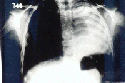
chest x-ray examination reveals opacity of the right hemithorax (Fig.
1). History reveals she sustained thoracic trauma after a
television
set fell over her chest two years previously. Since then, two
admissions
to different hospitals with diagnosis of pneumonia were reported. A
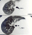
thoracic
CT-Scan revealed total atelectasis of the left lung (Fig.
2). Total rupture of the left main bronchus was detected during
bronchoscopic
examination.
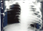
Resection of the
bronchial scar with bronchial anastomosis using absorbable synthetic
5-0
sutures resulted in progressive recovery of the affected lung of both
patients
(Fig. 3). They are both now free of symptoms, four
years and eighteen months after surgery respectively.
DISCUSSION
Although prompt
diagnosis and management of bronchial rupture secondary to blunt
thoracic
trauma is desirable, delayed surgical reconstruction of the main
bronchus
may be achieved without gross compromise of lung function (10).
Frequently
a "silent" rupture of the main bronchus may be misdiagnosed. Those
cases
demonstrates mild symptoms of respiratory insufficiency after trauma,
and
no pneumothorax or rib fractures at the radiological examination
of the chest. Days or even years may go by before the diagnosis is
made.
Partial or total lung atelectasis and extensive infiltrates in lung
parenchyma
diagnosed during routine radiological examination may be suspicious of
bronchial rupture after history of trauma. Thoracic CT-Scan or Magnetic
Resonance Imaging may help in diagnosis (11,12). With suspicion of
bronchial
rupture, bronchoscopy confirms the diagnosis (2,7,13,14). Both patients
had their diagnosis confirmed after fiberoptic bronchial examination
which
is of paramount importance in patients suffering blunt trauma having
symptoms
of respiratory distress, pleural air leaks, lobar atelectasis or
persistent
pneumothorax (7,9).
Immediate surgical
correction of bronchial rupture could reduce the incidence of late
complications
such as secondary tissue infection and persistent bronchial stenosis (
9, 15). In children, atelectatic lungs heal within a few weeks after
bronchial
reconstruction if no secondary infection is present (15). Bronchial
reconstruction
must always be attempted. Lung resection is restricted to those
patients
with tissue necrosis secondary to infection (9). Our two patients with
delayed diagnosis of bronchial rupture had no symptoms of infection.
They
were successfully managed with resection of the bronchial scar and
anastomosis
of the main bronchus with absorbable synthetic sutures. After
uneventful
surgical recovery they had follow-up bronchoscopic examination six
moths
later with total permeability of the main bronchus, a chest x-ray with
good ventilation of the affected lung and minimal elevation of the
diaphragmatic
dome.
BIBLIOGRAPHY
1.Hartley C, Morrit GN: Bronchial rupture secondary to blunt chest
trauma. Thorax 48:183-184, 1993
2. Goh SH, Tan SM, Chui P, Low BY: Traumatic bronchial rupture -
A case report. Ann Acad Med Singapore 24: 883-886, 1995
3. Baumgartner F, Sheppard B, de Virgilio C, Esrig B, Harrier D,
Nelson RJ, Robertson, JN: Tracheal and main bronchial disruptions after
blunt chest trauma: presentation and management. Ann Thor Surg
50:569-574,
1990
4. Frimpong-Boateng K, Amoah AGB: Delayed diagnosis and repair of
total bronchial rupture: A report of two cases. East Afr Med J
74:114-115,
1994
5. Roux P, Fisher RM: Chest injuries in children: na analysis of
100 cases of blunt chest trauma from motor vehicles accidents. J
Pediatr
Surg 27:551-555, 1992
6. Lin MY, Wu MH, Chan CS, Lai WW, Chou NS, Tseng YL: Bronchial
rupture by blunt chest trauma. Ann Emerg Med 25:412-415, 1995
7. Amauchi W, Birolini D, Branco PD, de Oliveira MR: Injuries to
the tracheobronchial tree in closed trauma. Thorax 38: 923-928, 1983
8. Sirbu H, Herse B, Schorn B, Hüttermann U, Dlichau H:
Successful
surgery after complete disruption of the right bronchial system. Thorac
Cardiovasc Surgeon 43: 239-141, 1995
9. Jones WS, Mavroudis C, Richardson JD, Gray LA Jr, Howe WR:
Management
of tracheobronchial disruption resulting from blunt trauma. Surgery 95:
319-323, 1984
10. Taskinen SO, Salo JÁ, Haltunen PE, Sovijarvi AR:
Tracheobronchial
rupture due to blunt chest trauma: a follow-up study. Ann Thor Surg 48:
846-849, 1989
11. Huson H, Sais CJ, Amendola MA: Diagnosis of bronchial rupture
with MR imaging. J Magn Reson Imaging 3: 919-920, 1993
12. Wan YL, Tsai KT, Yeow KM, Tan CF, Wong HF: CT findings of
bronchial
transection. Am J Emerg Med 15:176-177, 1997
13.Parat S, Bidat E, Chevallier B, Lacombe P, Azoulay R, Legardere
B, Bouledroua MS: Late diagnosis of a bronchial fracture after thoracic
injury. Ann Pediatr (Paris) 36:205-207, 1989
14. Rossbach MM, Johnson SB, Gomez MA, Sako EY, Miller OL, Calhoonn
JN: Management of major thacheobronchial injuries: a 28-year
experience.
Ann Thorac Surg 65:182-186, 1998
15. Gaebler C, Mueller M, Schramm W, Eckesberger F, Vecsei V:
Tracheobronchial
ruptures in children. Am J Emerg Med 14: 279-284, 1996
VENTILATORY STRATEGIES
TO PREVENT LUNG INJURY IN THE NEWBORN
Ramzi A.
Kilani, MD, FAAP
Assistant Professor of Pediatrics
Washington University School of Medicine
Division of Newborn Medicine
Ventilator-induced lung injury remains an important cause of morbidity and mortality in neonates requiring assisted mechanical ventilation. Historically, neonatal ventilation began as pressure-limited. Pressure, but not volume, was controlled. Hence, the term baro (airway pressure) trauma (lung injury). Animal studies showed, however, that changes in lung volume, and not pressure, caused lung injury [1]. This has led to a new term, volutrauma. Changes in pressure produce changes in volume as reflected by the compliance of the respiratory system.
Once the uninjured lung is recruited, it tends to stay open during exhalation. Surfactant and the structure of the lung and chest wall confer this deflation stability and define functional residual capacity (FRC). When FRC is optimized, end expiatory pressure can be decreased without a great loss of lung volume, as long as the pressure is not decreased below the critical closing pressure of the lung [2].
Therefore, ventilator induced lung injury sequence can occur at the beginning and end of each assisted breath. If inadequate numbers of alveoli were recruited in the previous breath, or if the positive end expiratory pressure was insufficient to maintain recruitment, atelectasis may occur, and can by itself propagate a lung injury sequence.
Once the airway epithelium and pulmonary capillary endothelium are injured, a lung injury sequence is initiated leading to alveolar capillary leak, surfactant disruption, inflammatory response and distal organ injury through efflux of inflammatory mediators from the alveolar space into the general circulation [3]. Oxidant injury may be another serious cause of lung injury. Immature and developing lungs are particularly susceptible to acquired injury.
An understanding of the basic pathophysiology of the underlying respiratory disorder is essential to optimize the ventilatory strategy. Aim for an adequate gas exchange without injuring the lungs.
Low tidal volume ventilation strategy
Focus ventilatory strategies for CMV in infants on prevention of overdistention, use of relatively small tidal volumes, and maintenance of adequate FRC. Studies in healthy infants report tidal volumes in the range of 5-8 cc/kg. A large multicenter trial in adults with acute RDS demonstrated that small tidal volumes (6 vs 12 mL/kg) and permissive hypercapnia significantly reduced mortality rates and ventilatory needs [4]. Insufficient data are available to recommend a specific size of tidal volume in infants with RDS who can be ventilated with even lower tidal volumes of 4-6 cc/kg. In infants with severe pulmonary disease, ventilate with small tidal volumes, because lung heterogeneity and unexpanded alveoli lead to overdistention and injury of the most compliant alveoli if a normal tidal volume is used.
Permissive hypercapnia strategy (PHS)
Permissive hypercapnia, or controlled mechanical hypoventilation, is a strategy for the treatment of patients receiving ventilatory assistance. When using this strategy, priority is given to the prevention or limitation of over ventilation rather than maintenance of normal blood gases and the high alveolar ventilation that frequently is used. Respiratory acidosis and alveolar hypoventilation may be an acceptable price for the prevention of pulmonary volutrauma. Thus, ventilatory strategies, which tolerate mild hypercapnia (PaCO2: 45-55 mm Hg) and/or prevent hypocapnia (particularly during the first days of life), may result in a reduced incidence and/or severity of lung injury [5, 6].
The limited number of randomized controlled trials that evaluated permissive hypercapnea in preterm infants have not demonstrated any overall benefit of ventilatory strategies that target high levels of PaCO2 (>55 mmHg). Further trials are needed to establish the safe, or ideal, range for PaCO2 in ventilated newborn infants [7].
Potential advantages of PHS are decreased volutrauma and lung injury, decrease duration of mechanical ventilation, reduced alveolar ventilation, reduced side effects of hypocapnea, and increased oxygen unloading. Potential disadvantages are cerebral vasodilation, hypoxemia, hyperkalemia, decreased oxygen uptake by hemoglobin, increased pulmonary vascular resistance, intraventricular hemorrhage in prematures, and retinopathy of prematurity.
Although, term and near term infants with respiratory failure and PPHN have been managed successfully with PaCO2 levels as high as 65 mm Hg, There are no published randomized controlled trials (RCTs) comparing outcomes in infants with PPHN using a strategy of permissive hypercapnea with one of hyperventilation or even one of achieving eucapnea.
Clearly, more RCTs are needed before permissive hypercapnea can be recommended routinely for preterm, near term, and term infants with respiratory disorders [8].
Strategies based on alternative modes of ventilation
Technologic advances have resulted in better ventilators. Patient-triggered ventilation and synchronized intermittent mandatory ventilation are being used increasingly in newborns. High-frequency ventilation is another mode of ventilation that may reduce lung injury and improve pulmonary outcomes, though available studies fail to demonstrate consistent benefits.
Patient-triggered ventilation
During conventional ventilation neonates are ventilated with continuous flow, pressure limited, time cycled ventilators. The introduction of triggered ventilation has been a marked improvement in neonatal ventilation. Various triggered ventilation modes have been developed for neonates: Synchronous Intermittent Mandatory Ventilation (SIMV), Assist/Control Ventilation (A/C), and Pressure Support Ventilation (PSV).
Consequences of Asynchrony
Asynchrony between spontaneous ventilation and mechanical breaths can be problematic. Asynchrony causing active expiration against ventilator inflation may occur irregularly or continuously, depending on the ventilator setting. Consequences of active expiration may be a decrease in tidal volume.
Synchronized intermittent mandatory ventilation
This mode of ventilation achieves synchrony between the patient and
the ventilator breaths. Synchrony may be achieved by adapting
ventilator
settings to the spontaneous breathing pattern, by using triggered
ventilation
modes, or by using heavy sedation or paralysis. Most Neonatologists do
not adapt the practice of regular paralysis in infants with active
expiration,
because paralysis has some disadvantages. Sedation is more often used
but
also shows some disadvantages.
Synchrony can sometimes be achieved by nearly matching the ventilator
frequency to the spontaneous respiratory rate or by simply ventilating
at relatively high rates (60-120 min). An alternative approach is to
detect
the infant’s inspiratory effort and use it to trigger positive
pressure
inflation (triggered ventilation).
Patient triggered ventilation (PTV)
The most frequently used ventilators in newborns are time-triggered at a preset frequency, but because of the available bias flow, the patient also can take spontaneous breaths. In contrast, patient-triggered ventilation (PTV), which also is called assist/control, uses spontaneous respiratory effort to trigger the ventilator. During PTV, changes in airway flow or pressure, chest wall or abdominal movements, or esophageal pressure changes are used as an indicator of the onset of the inspiratory effort. Once the ventilator detects inspiratory effort, it delivers a ventilator breath at predetermined settings (PIP, inspiratory duration, flow).
Despite short-term benefit, large randomized controlled trials report that patient-triggered ventilation does not improve long-term outcomes in infants with RDS [9], although' compared to conventional mechanical ventilation (CMV) PTV has been shown to improve tidal volume, oxygenation and reduce blood pressure fluctuations. PTV compared to SIMV was associated with a trend to a shorter duration of weaning [9].
High-frequency ventilation
High-frequency oscillatory ventilation (HFOV) delivers very small tidal volumes (even smaller than dead space) generated by an oscillatory piston or diaphragm, at extremely high frequencies, superimposed on a variable mean airway pressure. Oscillatory ventilation is unique because exhalation is generated actively, as opposed to other forms of HFV (HFJV and HFII), in which exhalation is passive. Although, multiple studies in animals suggested advantage of HFOV for supporting immature or injured lungs, clinical trials of the routine use of HFV in infants have yielded mixed results [10]. When a high volume strategy (HVS) was used for HFOV, it was associated with significantly lower rates of CLD or death in survivors at 28-30 days and oxygen use at 36-37 weeks postmenstrual age or discharge. There were no overall differences in the rates of IVH or PVL [10].
Recently, Johnson et al (UK) [11] and Courtney et al (USA) [12] published their RCTs comparing HFOV and CMV in preterm infants. Both included infants who were at high risk (GA 23-28 weeks, and BW 601-1200 respectively). The results of the study by Johnson et al. showed no difference between the modes of ventilation in the combined primary outcome of death or chronic lung disease. In contrast, Courtney et al. found that HFOV conferred a small but significant benefit in survival without CLD (56 percent vs. 47 percent), and infants assigned to HFOV were successfully extubated one week earlier. The trial by Johnson received some criticism, while the trial by Courtney et al was conducted under rigorously controlled conditions with well-defined protocols for the management of HFOV [13]. It seems that HFOV administered according to strict protocols may be preferable in the most experienced centers. However, for most preterm infants, CMV with low tidal volumes and reasonable ventilation goals remains the appropriate choice.
PATHOPHYSIOLOGY-BASED VENTILATORY STRATEGIES
Respiratory distress syndrome
RDS is characterized by low compliance and low FRC. An optimal
conventional
ventilation strategy may include conservative indications for
conventional
ventilation, the lowest PIP and tidal volume required, modest
PEEP
(3-5 cm H20), permissive hypercapnia (PaCO2 45-55 mm Hg), judicious use
of sedation/paralysis, and aggressive weaning.
Chronic lung disease
BPD usually has heterogeneous time constants among lung areas. Resistance may be increased markedly, and frequent exacerbations may occur. A higher PEEP (4-6 cm H20) often is used, and longer inspiratory and expiratory times with low rates are preferred. The infants’ additional spontaneous breaths can be supported by pressure support (PS) mode. PS effects an endurance training of respiratory muscles and reduces the work of breathing. Hypercarbia with compensated respiratory acidosis often is tolerated to avoid lung injury secondary to aggressive mechanical ventilation.
Persistent pulmonary hypertension of the newborn
Persistent pulmonary hypertension of the newborn may be primary or associated with aspiration syndrome, prolonged intrauterine hypoxia, congenital diaphragmatic hernia, or other causes. Ventilatory treatment of infants often is controversial and varies markedly among centers. In general, adjust FiO2 to maintain PaO2 at 80-100 mm Hg to minimize hypoxia-mediated pulmonary vasoconstriction; adjust ventilatory rates and pressures to maintain an arterial pH of 7.45-7.55 (sometimes combined with bicarbonate infusion). Take care to prevent extremely low PaCO2 (<20 mm Hg), which can cause cerebral vasoconstriction and subsequent neurologic injury. Permissive hypercapnea is considered investigational. Addition of inhaled nitric oxide to conventional ventilation reduces the need for extracorporeal membrane oxygenation. Furthermore, the use of HFV to further optimize delivery of inhaled nitric oxide, as a result of its ability to improve lung inflation, has been demonstrated [14].
Congenital Diaphragmatic hernia
An approach that challenged conventional wisdom in the context of a care strategy based on permissive hypercapnea/spontaneous respiration/elective repair has been anecdotally reported by different groups, and demonstrated improved survival. In this approach, lower PaO2 or oxygen saturation (preductal saturations of 75-85% in the first 4 hours of life and > 90% subsequently), higher PaCO2 (60-65 torr or less) and lower PH (> 7.20-7.25) were accepted. The PIP was limited to 24-30 and PEEP to 5 cm H2O. Ventilator adjustments were based on maintaining preductal saturation of 90% or greater with no evidence of evolving metabolic acidosis. Such an approach, although promising, need to be tested in prospective trials to determine the most appropriate management of the infant with CDH [15, 16, 17].
Abdominal Distension
Increased intra-abdominal pressure results in upward pressure on the diaphragm, reduces diaphragmatic excursion, and results in decreased compliance of the respiratory system in newborns with acute intra-abdominal disease, such as necrotizing enterocolitis, bowel obstruction, or postoperatively in infants with gastroschisis, omphalocele, or diaphragmatic hernia. Large tidal volume ventilation further exacerbates the hemodynamic compromise normally caused by positive pressure ventilation. Therefore, a strategy of using small tidal volumes at a higher respiratory rate and adequate PEEP to prevent end expiratory atelectasis seems reasonable. The use of both HFOV and HFJV have been documented to improve ventilation and hemodynamic variables [18].
Summary
Sound application of the basic concepts of gas exchange, and pulmonary mechanics is necessary to optimize mechanical ventilation. Employment of pathophysiology-based ventilatory strategies to prevent lung injury, and alternative modes of ventilation should result in further improvement in neonatal outcomes.
References:
1. Dreyfuss D, Saumon G: Ventilator-induced lung injury: Lessons
from
experimental studies. Am J Respir Crit Care Med 157:294, 1998
2. Hamilton PP, Onayemi A, Smyth JA, et al: Comparison of conventional
and high-frequency ventilation: Oxygenation and lung pathology. J Appl
Physiol 55:131, 1983
3. Auten RL, Vozzelli M, Clark RH: Volutrauma; What is it, and how
do we avoid it? Clinics in Perinatology; vol 28 (3); 2001, 505-509
4. The Acute Respiratory Distress Syndrome Network. Ventilation with
lower tidal volumes as compared with traditional tidal volumes for
acute
lung injury and the acute respiratory distress syndrome. N Engl J Med.
2000;342:1301-1308
5. Mariani G. Cifuentes J. Carlo WA. Randomized trial of permissive
hypercapnia in preterm infants. Pediatrics. 104(5 Pt 1):1082-8, 1999
Nov.
6. Gonzalo Mariani, Javier Cifuentes, and Waldemar A. Carlo. Minimal
ventilation to prevent bronchopulmonary dysplasia in
extremely-low-birth-weight
infants. The Journal of Pediatrics; Volume 141(3), 2002: 370-375
7. Woodgate PG. Davies MW. Permissive hypercapnia for the prevention
of morbidity and mortality in mechanically ventilated newborn infants
(Cochrane
Review). In: The Cochrane Library, Issue 3 2002. Oxford: Update
Software.
8. Ambalavanan N, Carlo Waldemar: Hypocapnia and hypercapnia in
respiratory
management of newborn infants. Clinics in Perinatology; vol 28 (3);
2001,
517-531
9. Greenough A, Milner AD, Dimitriou G: Synchronized mechanical
ventilation
for respiratory support in newborn infants (Cochrane review). In: The
Cochrane
Library, Issue 2, 2000. Oxford: Update Software
10. Henderson-Smart DJ, Bhuta T, Cools F, Offringa M. Elective high
frequency oscillatory ventilation versus conventional ventilation for
acute
pulmonary dysfunction in preterm infants (Cochrane Review). In: The
Cochrane
Library, Issue 4, 2002. Oxford: Update Software.
11. Johnson AH, Peacock JL, Greenough A, Marlow N, Limb ES, Martson
L, and Calvert SA. High-frequency oscillatory ventilation for the
prevention
of chronic lung disease of prematurity. N Engl J Med; 347-9: 633-642
12. Courtney SE, Durand DJ, Asselin JM, Hudak ML, Aschner JL, and
Stoemaker
GT. High-frequency oscillatory ventilation versus conventional
mechanical
ventilation for very-low-birth-weight infants
13. Stark AR: High-frequency oscillatory ventilation to prevent
bronchpulmonary
dysplasia - Are we there yet? N Engl J Med, 2002; 347-9: 682-683
14. Kinsella JP, Truog WE, Walsh WF, et al: Randomized, multicenter
trial of inhaled nitric oxide and high-frequency oscillatory
ventilation
in severe, persistent pulmonary hypertension of the newborn. J Pediatr
131:55-62, 1997
15. Boloker J. Bateman DA. Wung JT. Stolar CJ. Congenital diaphragmatic
hernia in 120 infants treated consecutively with permissive
hypercapnea/spontaneous
respiration/elective repair. Journal of Pediatric Surgery.
37(3):357-66,
2002 Mar.
16. Finer NN. Tierney A. Etches PC. Peliowski A. Ainsworth W.
Congenital
diaphragmatic hernia: Developing a protocolized approach. Journal
of Pediatric Surgery. 33(9):1331-1337, 1998
17. Wilson JM. Lund DP. Lillehei CW. Vacanti JP. Congenital
diaphragmatic
hernia-A tale of two cities: The Boston Experience. Journal of
Pediatric
Surgery. 32(3):401-405, 1997
18. Fok TF, Ng PC, Wong W, et al: High-frequency oscillatory
ventilation
in infants with increased intra-abdominal pressure. Arch Dis Child 76:
F123-125, 1997][keszler M, Jennings LL: High-frequency jet ventilation
in infants with decreased chest wall compliance. Pediatr Res 41:257 A,
1997
THE INCIDENCE OF INGUINAL
HERNIA
ASSOCIATED WITH UNDESCENDED TESTIS
*Naguib N.N., Samuel D.G., Izzidien A.Y.
Department of Surgery, Prince Charles Hospital.
Merthyr Tydfil, CF47 9DT.
*Corresponding author: nadernaguib71@yahoo.com
Introduction
In the eighth week of intrauterine life, the testes arise in the abdominal cavity and descend through the inguinal canal to the scrotum in the third trimester.¹ Testicular descent is a dynamic process, and about 5% of full term infants (and about 25% of premature infants) will not have a descended testis at birth.² 1.5% of boys at 3 months of age will still have undescended testis.³ Cryptorchidism and congenital inguinal hernia commonly co-exist, with the principal defect being a patent process vaginalis.?
Aim
In this study, we aim to assess the incidence of inguinal hernia /
patent process vaginalis in association with undescended testes.
Patients and Methods
We conducted a retrospective and prospective study, of 150 pediatric
patients (<16 years old) over a 5 year period, who had a first stage
orchidopexy at a district general hospital in Wales. Patients’
operative
notes were reviewed to record the procedure performed, location of the
undescended testis, whether unilateral or bilateral, and the presence
of
associated inguinal hernia / patent process vaginalis.
Results
Over a period of 5 years, 150 pediatric patients had 212 first stage
orchidopexy procedures for undescended testes. The age range was 1
– 15
years old with mean age 5.7 and median age of 8. Out of the 150
patients,
62 (41.3%) patients had bilateral procedure, 54 (36%) patients had
right
side orchidopexy and 34 (22.7%) patients had left side orchidopexy.
The majority of the testes were found in the inguinal canal (158/212
= 74.5 %), 32 (15%) testes were present at the superficial inguinal
ring
and 16 (7.5%) were at the deep inguinal ring. Only 5 testes (2.4%) were
intra-abdominal and 1 testis was ectopic in the superficial inguinal
pouch.
Out of the 62 bilateral orchidopexies, 57 (92%) patients had mirror
image testes, lying in the same site on the contra lateral side, while
5 (8%) patients had the two testes lying at 2 different levels.
All cases had exploration for the presence of inguinal hernia / patent
process vaginalis which was repaired, if found, at the same setting.
The
overall incidence of inguinal hernia / patent process vaginalis
associated
with undescended testis was 91% (193/212). The incidences were 93.5%
(58/62),
91% (49/54) and 85% (29/34) associated with bilateral, right and left
undescended
testes, respectively.
Only one of the 58 patients with bilateral undescended testes and
inguinal
hernia / patent process vaginalis had a unilateral hernia on the right
side, the remaining 57 patients had bilateral inguinal hernias / patent
processes vaginalis.
There were no complications that occurred in our patient cohort as
a result of surgery and no patients have reported a recurrence of their
inguinal hernia so far.
Discussion
The occurrence of a congenital inguinal hernia and undescended testis
is related to descent of the testis into the scrotum. The testes
usually
reach their final destination in the scrotum by the third trimester. In
approximately 90% of infants, the processus vaginalis seals and becomes
a thin band of tissue without a lumen or opening?. Sacs occur in this
region
if this muscular obliteration is incomplete and allows for herniation
as
well as abnormal testicular descent and positioning due to defective
muscular
propulsion?.
Patients may present at any age with an undescended testis although
most studies advocate an early intervention within the first 1-2 years
of life. This is seen as beneficial in preserving the testicle as well
as reducing the risk of subfertility & malignant transformation
seen
in later life???.
The incidence of inguinal hernia / patent process vaginalis reported
in association with cryptorchidism varies in the literature.
Several
studies suggest that Cryptochidism is almost always associated with an
indirect inguinal hernia???. In a study by Al-Abbadi and Smadi
conducted
on 37 children, the undescended testis associated with indirect
inguinal
hernia was reported in 75.68% ?. On the other hand, some studies
suggest
that only 20% of patients with undescended testes will have an inguinal
hernia ¹°?¹¹. Tanyel et al. and Davenport, clarified
that although the processus vaginalis is patent in boys with
undescended
testis, clinical inguinal hernia is only encountered in 10-15%
²??.
Our results confirm that inguinal hernias / patent process vaginalis
are a common co-occurrence in children who present with cryptorchidism.
And this seems the factor in preventing the normal descend of the
testis.
References
1. Russell RC, Norman SW, Christopher JK. The testis and scrotum. In:
Bailey and Loves Short practice of surgery. 23rd edn. London: Arnold:
2000.pp.1270-83.
2. Davenport M. ABC of General Paediatric Surgery: Inguinal
hernia,
Hydrocele, and the undescended testis. BMJ, March 1996;
312:
564 – 567.
3. John Radcliffe Cryptorchidism Study Group. Cryptorchidism: an
apparent
substantial increase since 1960. BMJ 1986; 293:1401-4.
4. Favorito, L.A. Costa, W.S. Sampaio, F.J. Relationship between the
persistence of the processus vaginalis and age in patients with
cryptorchidism.
Int Braz J Urol. 2005 Jan-Feb; 31(1):57-61.
5. American Pediatric Surgical Association.
http://www.eapsa.org/index.cfm
6. Tanyel , FC , Öcal T, Karaa?ao?lu E,
Büyükpamukçu
N. Individual and associated effects of length of inguinal canal and
caliber
of the sac on clinical outcome in children. J Pediatr Surg 2000; 35:
1165-1169.
7. Merck Manuals online medical library http://www.merck.com
8. Woodhouse CRJ. Late malignancy risk in urology. Br J Urol 1992;
70: 345-51. [Medline]
9. K. Al-Abbadi and S.A. Smadi. Genital abnormalities and groin hernias
in elementary-school children in Aqaba: an epidemiological study.
Eastern
Mediterranean Health Journal. Volume 6, Issue 2/3, 2000, Page 293-298.
10. John Radcliffe Cryptorchidism Study Group. Cryptorchidism:
an apparent substantial increase since 1960. BMJ 1986; 293: 1401-4.
11. Chilvers, C. Pike, M.C. Epidemiology of undescended testis.
In Oliver RTD, Blandy JP, Hope-Stone, HF. Eds. Urological and genital
cancer.
Oxford: Blackwell, 1989:306-21.
|
|
|
|
|
|
|
|
|
|
|
|
|
|
|
|
|
|