what is your diagnosis ?

 
CASE HISTORY 1:
Six day old baby girl born vaginally after 30 weeks gestation with persistently enlarged 
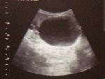
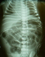
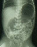
huge solitary gastric bubble, no distal air in the KUB (see adjacent 
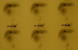
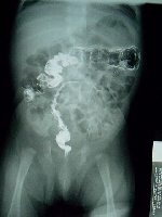
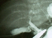
figure). 
She passed some meconium on the first day of life.  After 24 hrs of life she developed hypochloremia.
The UGIS shows a dilated stomach, a normal antrum with a cap and faint insinuation of a pyloric canal. 
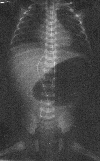
KUB

Click here for answer


CASE HISTORY 2:
14 year old black male with progressive dysphagia, regurgitation, weight
loss, and nocturnal cough. 

Barium swallow shows esophageal dilatation, motility alteration and a small caliber cardio-esophageal junction (see adjacent figure).


Barium Swallow

Click here for answer


CASE HISTORY 3:
4 month female child with a right upper quadrant abdominal mass and jaundice.

Ultrasound (US) and nuclear scintigram (HIDA) are included.


US

HIDA

Click here for answer


CASE HISTORY 4:
Baby girl born prematurely with the adjacent abdominal wall defect, interrupted dilated small bowel with associated micro-ileum and micro-colon.

Click here for answer


CASE HISTORY 5:
Baby boy born at term with the adjacent abdominal wall defect. 

Click here for answer


CASE HISTORY 6:
6 year-old-female with two month history of left upper quadrant abdominal pain, early satiety and non-bilious vomiting. Normal chemistry and amylase/lipase. CT Scan shows a cystic mass behind the stomach.

Click here for answer


CASE HISTORY 7:
2 weeks-old female patient born to a diabetic mother develops abdominal distension. Barium enema (see figure) shows a thin left colon with a transitional zone at the splenic flexure. Ganglion cells are present on suction rectal biopsy. 

Click here for answer


CASE HISTORY 8:
A 13 year-old female presented with primary amenorrhea, low abdominal pain and a palpable movable pelvic mass. CT shows multiple cystic cavities in the pelvis.

Physical Exam

Pelvic CT Scan

Click here for answer


CASE HISTORY 9:
48-hours-old term female develops abdominal distension, obstipation and bilious vomiting. Rectal exam reveals a WHITE meconium plug. Simple abdominal films demonstrate multiple dilated small bowel loops. At surgery there is a transitional zone 23 cm proximal to ileocecal valve with the proximal dilated ileum filled with tenacious neconium and distal meconium pellets.

KUB

Operative findings

Click here for answer


CASE HISTORY 10:
3 months-old-male infant with persistent bilious vomiting. Simple abdominal film is included. 

KUB

Click here for answer


CASE HISTORY 11:
3 months prematurely born (33 weeks gestational age) male child with history of hyaline membrane disease, pneumothorax, mechanical ventilation. convulsions, prolonged TPN and medically-managed NEC. While at NICU he develops insidious cholestatic jaundice (total bile fluctuating between 5 and 9 mg% with 70-80% conjugated portion). Elevated GGT and LFT's, normal alpha-1-antitrypsin levels and normal TORCH. HIDA scan under phenobarbital induction shows adequate liver uptake with no hepato-biliary excretion in 24 hrs films. Mini-laparotomy reveals a cholestatic liver, cholangiogram shows patent gallbladder, cystic duct and common bile duct with diminutive intrahepatic bile ducts (see figure). 

What is your diagnosis ?


CASE HISTORY 12:
Newborn female born with the congenital anomaly depicted in the adjacent picture.

What is your diagnosis?


Home
Table
Index
Past
Review
Submit
Techniques
Editor
Handbook
Articles
Download
UPH
Journal Club
WWW
Meetings
Videos
Created February 1, 1998
Last updated: May 2024